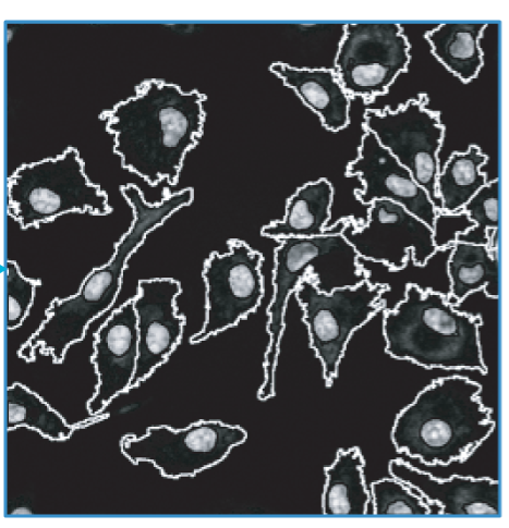
High-throughput cellular imaging and microfluidic technologies are enabling phenotypic measurements on single-cell and population-wide scales. The extraction of information from such imaging data is necessary for establishing the relationships between the behavior of molecular networks in cells and quantitative phenotypic features of cells and tissues. Image processing and analysis methods can help us detect, count and describe the shapes of subcellular and multicellular structures and track molecules or cells over time.
A. Fedorov, W. J. R. Longabaugh, D. Pot, D. A. Clunie, S. Pieper, H. J. W. L. Aerts, A. Homeyer, R. Lewis, A. Akbarzadeh, D. Bontempi, W. Clifford, M. D. Herrmann, H. Höfener, I. Octaviano, C. Osborne, S. Paquette, J. Petts, D. Punzo, M. Reyes, D. P. Schacherer, M. Tian, G. White, E. Ziegler, I. Shmulevich, T. Pihl, U. Wagner, K. Farahani, and R. Kikinis, “NCI imaging Data Commons,” Cancer Research, Vol. 81, No. 16, pp. 4188–4193, 2021.
J. Saltz, R. Gupta, L. Hou, T. Kurc, P. Singh, V. Nguyen, D. Samaras, K. Shroyer, T. Zhao, R. Batiste, J. Van Arnam, Cancer Genome Atlas Research Network, I. Shmulevich, A. Rao, A. Lazar, A. Sharma, V. Thorsson, “Spatial Organization and Molecular Correlation of Tumor-Infiltrating Lymphocytes Using Deep Learning on Pathology Images“, Cell Reports, Vol. 23, No. 1, pp. 181-193.e7, 2018.
P. Ruusuvuori, Jake Lin, A. C. Scott, Z. Tan, S. Sorsa, A. Kallio, M. Nykter, O. Yli-Harja, I. Shmulevich, A. M. Dudley, “Quantitative analysis of colony morphology in yeast,” BioTechniques, Vol. 56, No. 1, pp. 18–27, 2014.
D. Falconnet, A. Niemistö, R. J. Taylor, M. Ricicova, T. Galitski, I. Shmulevich, C. L. Hansen, “High-throughput tracking of single yeast cells in a microfluidic imaging matrix,” Lab on a Chip, Vol. 11, pp. 466-473, 2011.
R. A. Saleem, R. Long-O’Donnell, D. J. Dilworth, A. M. Armstrong, A. P. Jamakhandi, Y. Wan, T. A. Knijnenburg, A. Niemistö, J. Boyle, R. A. Rachubinski, I. Shmulevich, J. D. Aitchison, “Genome-Wide Analysis of Effectors of Peroxisome Biogenesis,” PLoS ONE, Vol. 5, No. 8, e11953, 2010.
J. Selinummi, P. Ruusuvuori, I. Podolsky, A. Ozinsky, E. Gold, O. Yli-Harja, A. Aderem, I. Shmulevich, “Bright Field Microscopy as an Alternative to Whole Cell Fluorescence in Automated Analysis of Macrophage Images ,” PLoS ONE, Vol. 4, No. 10, 2009.
R. J. Taylor, D. Falconnet, A. Niemistö, S. A. Ramsey, S. Prinz, I. Shmulevich, T. Galitski, C. L. Hansen, “Dynamic analysis of MAPK signaling using a high-throughput microfluidic single-cell imaging platform,” Proceedings of the National Academy of Sciences of the USA, Vol. 106, No. 10, pp. 3758-3763, 2009.
J. Selinummi, A. Niemistö, R. Saleem, G. W. Carter, J. Aitchison, O. Yli-Harja, I. Shmulevich, and J. Boyle, “A Case Study on 3-D Reconstruction and Shape Description of Peroxisomes in Yeast, ” IEEE International Conference on Signal Processing and Communication (ICSPC07), Dubai, United Arab Emirates (UAE), November 24-27, 2007.
A. Niemistö, T. Korpelainen, R. Saleem, O. Yli-Harja, J. Aitchison, I. Shmulevich, “A K-Means Segmentation Method for Finding 2-D Object Areas Based on 3-D Image Stacks Obtained by Confocal Microscopy,” 29th International Conference of the IEEE Engineering in Medicine and Biology Society, Lyon, France, August 23-26, 2007.
A. Niemistö, J. Selinummi, R. Saleem, I. Shmulevich, J. Aitchison, O. Yli-Harja, “Extraction of the number of peroxisomes in yeast cells by automated image analysis,” The 28th Annual International Conference of the IEEE Engineering in Medicine and Biology Society, New York, New York, August 30 – September 3, 2006.
A. Niemistö, V. Dunmire, O. Yli-Harja, W. Zhang, I. Shmulevich, “Robust quantification of in vitro angiogenesis through image analysis,” IEEE Transactions on Medical Imaging, Vol. 24, No. 4, pp. 549-553, 2005.
A. Niemistö, V. Dunmire, O. Yli-Harja, W. Zhang, I. Shmulevich, “Analysis of angiogenesis using in vitro experiments and stochastic growth models,” Physical Review E, Vol. 72, 062902, 2005.
A. Niemistö, I. Shmulevich, O. Yli-Harja, L. Chirieac, S. R. Hamilton, “Automated Quantification of Lymph Node Size and Number in Surgical Specimens of Stage II Colorectal Cancer,” 27th Annual International Conference of the IEEE Engineering in Medicine and Biology Society, Shanghai, China, September 1-4, 2005, pp. 6313 – 6316.
A. Niemistö, L. Hu, O. Yli-Harja, W. Zhang, I. Shmulevich, “Quantification of in vitro cell invasion through image analysis,” in Proceedings of the 26th Annual International Conference of the IEEE Engineering in Medicine and Biology Society (EMBS’04), San Francisco, California, USA, Sep. 1-5, 2004, pp. 1703-1706.


 shmulevich.isbscience.org/research/image-analysis/